The Ultimate Guide to Thermography: Decoding the Body’s Silent Signals
.
Thermography detects heat on the skin’s surface using an infrared camera and is a safe, painless, and cost effective screening tool used for prevention.
The body constantly speaks to us, sending signals that alert us to problems needing our attention. One of those signals is increased heat—which can point to ailments in their early stages.
Thermography is a unique screening technique that translates the body’s thermal signals into a colorful map—offering a powerful tool for timely detection and prevention of various health conditions.
.
How Thermography Works
Thermography, also known as digital infrared thermal imaging (DITI), is a non-invasive screening test that uses an infrared camera to detect subtle temperature changes on the skin’s surface. These changes can provide clues about what’s happening inside the body, revealing factors such as inflammation, hormonal imbalances, and lymphatic congestion, which can lead to a buildup of fluid in the body’s tissues.Dr. Christine Horner, a board-certified general and plastic surgeon, award-winning author, natural health expert, and founder of a thermography practice in Southern California, spoke to The Epoch Times about thermography and how it works.
“It’s measuring the surface temperature of the skin, and then the software program will assign a different color to every temperature—and that’s what creates our image on our computer screen,” she said. “So, in essence, whatever area of the body that we’re looking at, you can think of it as a geographical temperature map,” she added.
The procedure is simple, safe, painless, and relatively low-cost. Unlike other imaging methods, there’s no compression or radiation involved, and the camera never touches the patient.
.
Thermography vs. Traditional Imaging
Thermography differs from tests like mammography or ultrasound in that it’s a test of physiology or function, rather than anatomy or structure. Combining both types of tests can provide more comprehensive information about changes in the body and potential pathologies.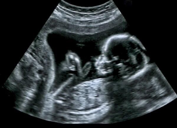
Ultrasound of a baby in a mother's womb. GagliardiPhotography/Shutterstock
.
Thermography Basics
Thermography’s effectiveness is rooted in its connection to the sympathetic nervous system, which controls the body’s fight or flight response and innervates the skin, according to Horner.“Whenever there’s any kind of underlying imbalances, dysfunctions, injuries, infections—anything like that going on—the sympathetic nervous system is going to cause the overlying skin temperature to change,” she said.
.
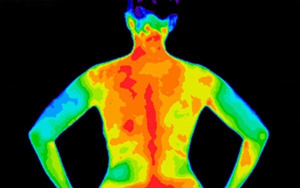
Anita van den Broek/Shutterstock
.
Thermal patterns are also unique to individuals—like fingerprints—and can change over time, Shawn Seuferer, a certified clinical thermographer, told The Epoch Times. There are specific thermal and colored patterns indicative of dysfunction depending on where they are on the body, she added.
“It’s not that specific where it can be diagnostic, but there are indicators which would correspond to, say, a dysfunction or a blockage or inflammation or hormone imbalance,” she said.
What Thermography Can Detect
Thermography can help visualize a wide range of dysfunctions in the body, according to Horner’s website, including:- Unexplained pain and inflammation
- Digestive issues (liver, stomach, pancreas, spleen, intestines)
- Leaky gut
- Thyroid function challenges
- Immune imbalance
- Dental and sinus issues
- TMJ (disorders of the temporomandibular joint in the jaw)
- Neck and spinal problems
- Breast health
- Breast risk health assessment
- Lymphatic congestion
- Hormone imbalance
- Injury assessment and monitoring
- Musculoskeletal issues
.
Creating a Baseline–Why That’s Important
One of thermography’s key benefits is its ability to create a baseline for each patient through annual scans. Each patient’s infrared breast scan is unique, like a fingerprint. Any changes in this pattern over time can signal a potential problem.In cancer-free patients, the results can help assess future cancer risk.
Horner suggests women begin having regular scans in their 20s. “When we have a woman who has been a regular patient, and she comes in this year, and there’s an obvious difference compared to last year, she’s got that much-advanced warning now that there is a change, or there’s something going on—this gives her more options,” Seuferer said.
Thermography is a valuable tool for men as well, Seuferer added. “The images present indicators for inflammatory, neurological, vascular/lymphatic dysfunction applicable for a number of screenings for men and women: disc disease, nerve damage, undetected diabetic lesions, arthritis, fibromyalgia, Raynaud’s Syndrome, complex regional pain syndrome (CRPS), sprain/strain, vascular disease, undetected dental issues and TMJ, stroke screening, whiplash, inflammatory pain, allergy testing, etc.,” she said.
.
What to Do When Problems Arise
When changes are detected during thermographic imaging, further steps are typically recommended since thermography itself is not a diagnostic test.Further testing is the next step once anomalies are detected, Seuferer said. These include another thermogram to confirm, an ultrasound, and then, if there is still a question, more invasive tests like mammograms or an MRI.
“Truly, the only diagnostic tool is a biopsy, and not everybody wants to jump into that,” she added. “Some people say, just do it, cut it out, and I want to be done with it. But not everybody feels that way,” she noted.
Horner has a similar approach.
.
Factors Affecting Thermography
Several factors can affect a thermal image.- Recent exercise
- Consumption of caffeine, nicotine, or alcohol
- Certain medications and supplements
- Recent surgeries or dental work
- Tattoos and scarring
- Sunburns
- Room temperature
Activities, foods, and beverages that increase body temperature should be limited before a scan as they can also affect results, Horner added. “Exercise is not recommended beforehand, too, because, again, we’re increasing blood flow, and that is going to cause more heat in the body and in certain patterns,” she said. “Caffeinated products like coffees you shouldn’t have for at least two hours beforehand, not smoking, because of the nicotine in it, and no alcohol,” she added.
Even a sunburn can have an effect, and Horner recommends not tanning or having a sunburn for at least one week prior to your appointment.
The room temperature is also adjusted to be slightly cool for the best results.
.
Breast Thermography: A Preventative Tool
Although you can use thermography anywhere on the body, breast scans are commonly used to track any changes that may occur over time. Breast thermography is also used to detect changes associated with cancer or precancerous cell growth.“We’re not looking for breast cancer; we’re looking for physiological changes that can sometimes occur years before somebody would develop a breast cancer, and usually—fortunately—we can pick things up at such an early stage of just imbalances that it’s easily reversible with simple diet and lifestyle techniques,” Horner said. “So we can truly use it as a preventative tool, and that’s the reason I’m so passionate about it,” she added.
.
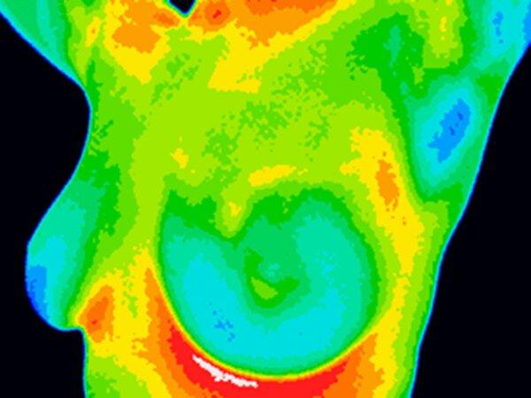
Anita van den Broek/Shutterstock
.
“The concept behind this test is that as cancer cells multiply, they need more oxygen-rich blood for growth. As there is an increase in blood flow to the tumor, the temperature around the tumor also increases. Malignant cells discharge nitric oxide into the bloodstream and cause impairment in the microcirculation. This released nitric oxide, along with the active growth of the cancerous cells, increases blood circulation and temperature in that particular region. Therefore, evaluating these differences in temperature leads to the detection of the malignant region in the breast.”
According to the International Academy of Clinical Thermology, studies support using thermography in the detection of breast cancer.
They say an abnormal infrared image is the most important predictor of developing breast cancer and is eight times more significant than a family history of the disease.
Thermography vs. Mammography
Breast cancer is the second leading cause of death in women. On average, a woman in the United States has a 13 percent chance of being diagnosed with breast cancer at some point in her life. This equates to a one in eight likelihood of developing the disease. About 60 percent of breast cancer deaths are due to delays in diagnosis, making early detection critical. Two main options exist: mammography and thermography.Mammography: ‘The Gold Standard’
Mammography has been deemed the gold standard for breast cancer detection for decades. It uses low-dose X-rays to identify abnormalities within the breast tissue. While it can detect breast cancer, it has risks and limitations:- False negatives: Dense breast tissue can obscure tumors. Mammography fails to detect one in five breast cancers, and false-negative results occur in one in eight cases, according to a 2022 review. Additionally, mammograms may identify non-cancerous anomalies, which often lead to unnecessary follow-ups, including additional mammograms and biopsies. These further procedures can cause unnecessary stress for both the body and the patient.
- Radiation exposure: Repeated mammograms expose women to low-dose radiation.
- Overdiagnosis: One systematic review estimated the breast cancer overdiagnosis rate at 52 percent. The review concluded that one in three breast cancers detected in women offered a mammography is overdiagnosed.
.
Thermography: A Promising Alternative
Thermography uses infrared imaging to detect variations in breast tissue temperature. Recent advancements in technology have made it a more viable option. Potential benefits include:- Early detection: Thermography may detect precancerous changes earlier than mammograms.
- No radiation: Thermography is a non-invasive procedure with no radiation exposure.
- Suitable for dense breasts: Thermography works well for women with dense breasts where mammograms are less effective.
- Pregnant or breastfeeding women
- Women with breast implants or who have had mastectomies
- Women with dense breasts
- Younger women
- Women at higher risk of breast cancer and need screening more often
- Women concerned about radiation exposure and breast compression
Breast Density
One significant benefit of thermography is its effectiveness for women with dense breasts, which contain more fibrous and glandular tissue than fat.Breast density is classified into four levels, ranging from mostly fatty tissue to highly dense tissue, according to the non-profit group Are You Dense?, which raises awareness about the impact of breast density on cancer detection via mammograms. Approximately 40 percent of women have dense breasts, making it more challenging to detect breast cancer through mammography.
Limitations of Thermography
- Accuracy: While promising, research on thermography’s accuracy is ongoing.
- Interpretation: Requires a trained thermologist to interpret images accurately
- Cause of increased heat: A warm reading may not be cancer, requiring further investigation.
.
The Takeaway
When it comes to our health, knowing about all the options is critical to making informed decisions.In the present health care system, some tests and treatments are widely available and easily accessible, while other equally viable alternatives are overlooked.
If you want options other than the ones your physician is recommending, ask. If your questions are not well received, find a doctor willing to have those discussions. Seuferer recommends gathering a team of supportive health care practitioners, particularly when facing a health challenge.
“I tell my patients, you’re hiring your care team, so you have the opportunity to select who you want to work with and who you do not want to work with. And it takes a lot of strength for a patient to tell their doctor, ‘No, I’m not going to work with you. I’m doing something different.’ The pressure is so immense,” she said.
Experts recommend not relying solely on one test but using a combination of methods to track your health and investigate concerns.
“The screening can be used as a pre-hire health assessment, used in a court of law, can be used in research, in medical practices, etc. It sounds like a bit of a ”one-stop-shop,“ and it is not meant to be,” Seuferer said. “Thermography is to be used as an adjunct to other modalities to help with diagnoses and help monitor the progression of treatment or therapy. When incorporated as an early indicator, it can provide the patient evidence to engage earlier intervention and thus have a greater potential for a positive outcome of injuries and other conditions,” she added.
Ultimately, there are many ways to deal with any health issue—and how to proceed is up to you.
.
What's Your Reaction?
 Like
0
Like
0
 Dislike
0
Dislike
0
 Love
0
Love
0
 Funny
0
Funny
0
 Wow
0
Wow
0
 Sad
0
Sad
0
 Angry
0
Angry
0


Comments (0)