What is a CT scan? Types, Uses, Procedure, and Risks
Contributed by: Anjali Sharma Introduction Over the years various types of diagnostic techniques have been invented by doctors and other healthcare experts to examine the symptoms and organs of a person’s body one such technique is CT Scan. A thorough examination of your body’s tissues, blood vessels, and bones is necessary for some medical diseases. While X-rays and ultrasounds can offer some insight, a computed tomography (CT) scan is typically the next step when a more thorough picture is needed. In this article, we’ll examine a CT scan’s operation, usual applications, and methods of usage in more detail. What is a CT Scan? CT scan is the medical diagnostic process through which the doctor gets an inside view of your body. It takes photographs of your organs, bones, and other tissues using X-rays and a computer. It displays more information than a standard X-ray. A CT scan can be done for any part of your body. It doesn’t take long, and there is no discomfort. How Do CT Scanners Operate? During a CT scan, a specific area of your body is circled by a focused X-ray beam. This is a collection of pictures taken from many viewpoints. This data is used by a computer to produce a cross-sectional image. This two-dimensional (2D) scan displays a ‘slice’ of the interior of your body, similar to one piece in a loaf of bread. A number of slices are created by repeating this procedure. These scans are stacked on top of one another by computer to produce an intricate representation of your inside organs, bones, or blood arteries. For instance, a surgeon would use this kind of scan to examine a tumour from all angles in order to plan an operation. Types of CT Scans Following are some different types of CT scan techniques. CT Angiography A doctor may request a CT angiography, often known as an angiogram, to determine a patient’s risk of developing heart disease. The scan can assist medical professionals in identifying blood vessel problems like aneurysms or blockages. A healthcare provider injects dye into the blood vessels prior to the scan that makes the blood flow through the body visible more clearly. The blood arteries are then captured on film by a CT technician. CT Abdomen Scan A technologist will use a scanner to produce pictures of the intestines, colon, liver, spleen, and appendix during an abdominal CT scan. An abdominal scan may be prescribed by a doctor to look for malignancies, such as colon tumours, or to identify and rule out abscesses in the region, internal bleeding, or both. Medical experts occasionally utilise a CT scan of the bones in addition to using x-rays to identify fractures and other issues with the bones. If the results of a conventional x-ray are uncertain, a doctor would prescribe a CT scan since it can provide more details. The tendons and muscles that are close to the bones may be seen more clearly thanks to a bone CT scan. The detection of bone cancer may also be aided by a CT scan of the bones. Head CT Scan If a patient complains of inexplicable headaches or dizziness, a doctor may recommend a head CT scan. The technique can aid in the diagnosis of strokes or brain malignancies. Images of the brain and other parts of the head, such as the sinuses, are captured during a head CT. A head CT may be helpful for patients with persistent sinus difficulties to find out if the region is still inflamed. Chest CT Scan A CT scan of the chest and lungs can provide a doctor with precise pictures of a patient’s lungs. If a patient complains of chest discomfort or breathing difficulties, a doctor may request a scan. Doctors can use the pictures to identify diseases including lung cancer, pneumonia, TB, or an excessive amount of fluid in the lungs. Cardiac CT A chest region image is also taken during a cardiac CT scan. The heart, as opposed to the lungs, is what is important. A cardiac CT may be prescribed by a physician to look for issues with the aorta, heart valves, or other arteries. When a treatment, such as coronary artery bypass grafting, has been performed, a doctor may occasionally request a cardiac CT scan to monitor the patient’s condition. Neck CT Scan The region from the base of the skull to the top of the lungs is routinely imaged during a CT scan of the neck. Tumours or masses in the neck, on the tongue, on the vocal cords, or in the upper airway can be found and diagnosed with the scan. A neck CT scan can also be used by a doctor to find growths or other abnormalities on the thyroid gland or problems with the carotid artery. Pelvic CT Scan A pelvic CT scan will produce images of the region of the body between the hip bones. It can aid in the diagnosis of abnormalities in the male or female reproductive systems as well as the detection of bladder disorders

Introduction
Over the years various types of diagnostic techniques have been invented by doctors and other healthcare experts to examine the symptoms and organs of a person’s body one such technique is CT Scan.
A thorough examination of your body’s tissues, blood vessels, and bones is necessary for some medical diseases. While X-rays and ultrasounds can offer some insight, a computed tomography (CT) scan is typically the next step when a more thorough picture is needed.
In this article, we’ll examine a CT scan’s operation, usual applications, and methods of usage in more detail.
What is a CT Scan?
CT scan is the medical diagnostic process through which the doctor gets an inside view of your body.
It takes photographs of your organs, bones, and other tissues using X-rays and a computer. It displays more information than a standard X-ray.
A CT scan can be done for any part of your body. It doesn’t take long, and there is no discomfort.
How Do CT Scanners Operate?
During a CT scan, a specific area of your body is circled by a focused X-ray beam. This is a collection of pictures taken from many viewpoints. This data is used by a computer to produce a cross-sectional image. This two-dimensional (2D) scan displays a ‘slice’ of the interior of your body, similar to one piece in a loaf of bread.
A number of slices are created by repeating this procedure. These scans are stacked on top of one another by computer to produce an intricate representation of your inside organs, bones, or blood arteries. For instance, a surgeon would use this kind of scan to examine a tumour from all angles in order to plan an operation.
Types of CT Scans
Following are some different types of CT scan techniques.
CT Angiography
A doctor may request a CT angiography, often known as an angiogram, to determine a patient’s risk of developing heart disease. The scan can assist medical professionals in identifying blood vessel problems like aneurysms or blockages.
A healthcare provider injects dye into the blood vessels prior to the scan that makes the blood flow through the body visible more clearly. The blood arteries are then captured on film by a CT technician.
CT Abdomen Scan
A technologist will use a scanner to produce pictures of the intestines, colon, liver, spleen, and appendix during an abdominal CT scan. An abdominal scan may be prescribed by a doctor to look for malignancies, such as colon tumours, or to identify and rule out abscesses in the region, internal bleeding, or both.
Medical experts occasionally utilise a CT scan of the bones in addition to using x-rays to identify fractures and other issues with the bones. If the results of a conventional x-ray are uncertain, a doctor would prescribe a CT scan since it can provide more details.
The tendons and muscles that are close to the bones may be seen more clearly thanks to a bone CT scan. The detection of bone cancer may also be aided by a CT scan of the bones.
Head CT Scan
If a patient complains of inexplicable headaches or dizziness, a doctor may recommend a head CT scan. The technique can aid in the diagnosis of strokes or brain malignancies. Images of the brain and other parts of the head, such as the sinuses, are captured during a head CT.
A head CT may be helpful for patients with persistent sinus difficulties to find out if the region is still inflamed.
Chest CT Scan
A CT scan of the chest and lungs can provide a doctor with precise pictures of a patient’s lungs. If a patient complains of chest discomfort or breathing difficulties, a doctor may request a scan. Doctors can use the pictures to identify diseases including lung cancer, pneumonia, TB, or an excessive amount of fluid in the lungs.
Cardiac CT
A chest region image is also taken during a cardiac CT scan. The heart, as opposed to the lungs, is what is important. A cardiac CT may be prescribed by a physician to look for issues with the aorta, heart valves, or other arteries.
When a treatment, such as coronary artery bypass grafting, has been performed, a doctor may occasionally request a cardiac CT scan to monitor the patient’s condition.
Neck CT Scan
The region from the base of the skull to the top of the lungs is routinely imaged during a CT scan of the neck. Tumours or masses in the neck, on the tongue, on the vocal cords, or in the upper airway can be found and diagnosed with the scan. A neck CT scan can also be used by a doctor to find growths or other abnormalities on the thyroid gland or problems with the carotid artery.
Pelvic CT Scan
A pelvic CT scan will produce images of the region of the body between the hip bones. It can aid in the diagnosis of abnormalities in the male or female reproductive systems as well as the detection of bladder disorders such as tumours or bladder stones.
Kidney CT Scan
Finding and confirming the presence of kidney stones is a frequent cause of a CT scan of the kidneys. The scan can aid in locating malignancies, abscesses, and renal disease symptoms.
Spine CT Scan
The imagery of the skeletal spinal structure, the discs between the bones, and the soft tissue of the spinal column are all captured by a spinal CT scan. A CT scan of the spine can be used to identify herniated discs, diagnose injuries to the area, and examine the region prior to surgery.
A physician may occasionally use a spinal CT to assess the degree of bone loss brought on by osteoporosis in the region. Additionally, a CT scan of the spine might help with a biopsy or other techniques.
Uses of CT Scan
There are several reasons why doctors prescribe CT scans, including
- CT scans are able to identify malignancies and complicated bone fractures, among other joint and bone conditions.
- CT scans may detect conditions like cancer, heart disease, emphysema, or liver tumours and enable medical professionals to notice any changes in such conditions.
- They exhibit internal bleeding and wounds similar to those from auto accidents.
- A tumour, blood clot, surplus fluid, or infection may be located with their assistance. In order to direct treatment plans and operations like biopsies, surgeries, and radiation therapy, doctors employ them.
- To determine if certain therapies are effective, doctors might compare CT scans. For instance, repeated scans of a tumour over time might reveal how well either chemotherapy or radiation is working.
CT Scan procedures
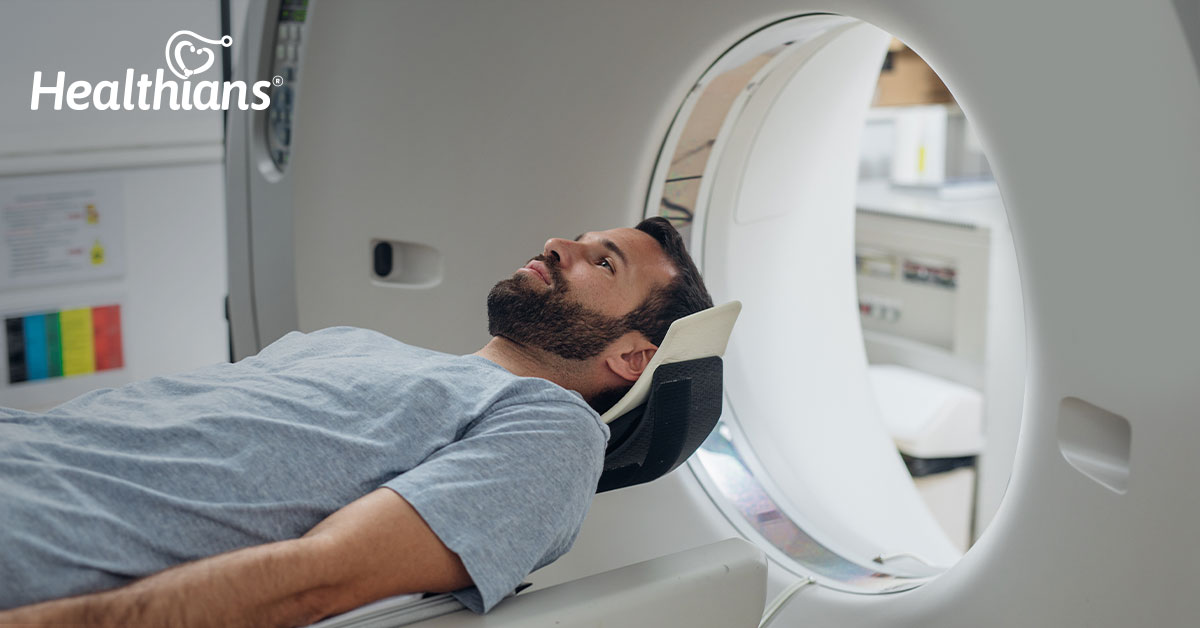
For a CT scan, the patient is asked to lie in a sleeping position. The scanner passes over the patient once they are lying down in the sleeping posture.
For optimal pictures created during the scan, the patients are asked to maintain their stillness and stability. The technicians relocate to the adjacent room for the duration of the process, although there is a facility for intercom contact between the technician and the patient.
The major bodily components are X-rayed repeatedly by the CT scanner. In order to acquire good insights, the photos are taken from various perspectives. In contrast to MRI equipment, the CT scan machine is not loud throughout the whole procedure.
Risks of CT Scan
CT scans are usually regarded as safe by medical professionals. Children’s CT scans are secure as well. Your CT technician could employ equipment designed with children in mind to minimise their radiation exposure to them.
Like other diagnostic procedures, CT scans create the picture using a tiny amount of ionising radiation. Among the dangers connected to CT scans are:
- Cancer risk: All radiation-based imaging procedures, like X-rays, slightly raise your chance of getting cancer. The change is too little to accurately measure.
- Allergic risks: Occasionally, persons may experience a mild or severe allergic response to the contrast agent.
Speak with your healthcare professional if you have any questions regarding the potential health hazards of CT scans. They will go through your concerns and aid you in making an informed decision.
Is CT Scan safe for a pregnant woman?
Inform the CT technician if you are or think you might be pregnant. The growing foetus may be exposed to radiation during pelvic and abdominal CT scans, but not to an extent that would be harmful. The foetus is not in any danger from CT scans in other sections of the body.
Final thoughts
The CT scan is a superior diagnostic tool. When your doctor requests a CT scan, you might be concerned. However, there is very little danger associated with this simple, safe examination. The benefit is that a CT scan can assist your doctors in correctly diagnosing a health issue and offering you the best course of therapy.
Any concerns that you might experience, including other testing possibilities, should be discussed with your health care professional.


