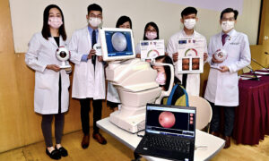
CUHK Medical Researchers Develop World’s First AI Retinal Imaging Analysis to Detect Alzheimer’s Disease
In Hong Kong, one in 10 elders aged 70 or more suffers from a cognitive disorder; more than half of the patients have Alzheimer’s disease. A medical team of the Chinese University of Hong Kong’s (CUHK) Faculty of Medicine has recently developed the world’s first artificial intelligence model using photographs of the fundus (back interior surface of the eye) alone to detect Alzheimer’s disease. [1] With a single retinal image scan, the system can detect Alzheimer’s disease with over 80 percent accuracy. The Automatic Retinal Image Analysis (ARIA-stroke) system is user-friendly, non-invasive, cost-effective, and helps to identify high-risk cases of recessive Alzheimer’s disease. [2] The gradual death of brain cells in patients with Alzheimer’s is caused by the excessive accumulation of abnormal substances in the brain, such as beta-like amyloid proteins and tangled nerve fibers. Clinical Professional Consultant Dr. Lisa Au Wing-chi at the Department of Medicine and Therapeutics, Faculty of Medicine, CUHK, expressed that it is difficult to accurately diagnose Alzheimer’s disease based on cognitive tests and brain structure imaging scans. The methods used for detecting amyloid proteins at the moment, such as a positive electron brain scan or extraction of cerebrospinal fluid through a vertebral puncture, are still uncommon and invasive. The human retina is an extension of the central nervous system. Therefore, non-invasive scanning methods such as ARIA-Stroke can reflect whether Alzheimer’s disease-related lesions are apparent in the cerebral blood vessels or the optic nerves. The CUHK clinical team developed and tested the ARIA-Stroke system using nearly 13,000 retinal image analyses from 648 patients with Alzheimer’s disease and 3,240 people with normal cognitive function. The experiment also confirmed that the accuracy of ARIA-Stroke reaches 84 percent, while the sensitivity and specificity are 93 percent and 82 percent, respectively. The accuracy remains between 80 to 92 percent when data from people of different ethnicities and countries are applied to the new ARIA-Stroke system. Professor Mok Hing-yiu of the Chinese University of Hong Kong, and Director of Therese Pei Fong Chow Research Centre for Prevention of Dementia, emphasized the system’s accuracy. The accuracy of the new ARIA-Stroke system is comparable to that of imagery examinations using magnetic resonance imaging, known as MRI. Additionally, the new system is user-friendly and non-invasive. ARIA-Stroke can potentially aid in diagnosing Alzheimer’s disease in clinics, as well as becoming a valuable tool for the community to screen for the disease. The team expects to incorporate the artificial intelligence system to identify high-risk patients of Alzheimer’s disease hidden in the community in the near future, so various preventive treatments can start as soon as possible to slow down patients’ cognitive degeneration and lessen brain damage. “Even if the tested patients suffer from ophthalmological diseases, such as macular lesions and glaucoma, ARIA-Stroke can still effectively detect retinal features associated with Alzheimer’s disease,” said Associate Professor Cheung Yim Lui at the CUHK Department of Ophthalmology and Visual Science. Cheung said that the CUHK Medical Centre is currently developing a comprehensive artificial intelligence system that will be able to detect Alzheimer’s disease by combining data on diseases relating to lesions of retinal blood vessels and nerves. Follow Follow

In Hong Kong, one in 10 elders aged 70 or more suffers from a cognitive disorder; more than half of the patients have Alzheimer’s disease.
A medical team of the Chinese University of Hong Kong’s (CUHK) Faculty of Medicine has recently developed the world’s first artificial intelligence model using photographs of the fundus (back interior surface of the eye) alone to detect Alzheimer’s disease. [1]
With a single retinal image scan, the system can detect Alzheimer’s disease with over 80 percent accuracy.
The Automatic Retinal Image Analysis (ARIA-stroke) system is user-friendly, non-invasive, cost-effective, and helps to identify high-risk cases of recessive Alzheimer’s disease. [2]
The gradual death of brain cells in patients with Alzheimer’s is caused by the excessive accumulation of abnormal substances in the brain, such as beta-like amyloid proteins and tangled nerve fibers.
Clinical Professional Consultant Dr. Lisa Au Wing-chi at the Department of Medicine and Therapeutics, Faculty of Medicine, CUHK, expressed that it is difficult to accurately diagnose Alzheimer’s disease based on cognitive tests and brain structure imaging scans.
The methods used for detecting amyloid proteins at the moment, such as a positive electron brain scan or extraction of cerebrospinal fluid through a vertebral puncture, are still uncommon and invasive.
The human retina is an extension of the central nervous system. Therefore, non-invasive scanning methods such as ARIA-Stroke can reflect whether Alzheimer’s disease-related lesions are apparent in the cerebral blood vessels or the optic nerves.
The CUHK clinical team developed and tested the ARIA-Stroke system using nearly 13,000 retinal image analyses from 648 patients with Alzheimer’s disease and 3,240 people with normal cognitive function. The experiment also confirmed that the accuracy of ARIA-Stroke reaches 84 percent, while the sensitivity and specificity are 93 percent and 82 percent, respectively.
The accuracy remains between 80 to 92 percent when data from people of different ethnicities and countries are applied to the new ARIA-Stroke system.
Professor Mok Hing-yiu of the Chinese University of Hong Kong, and Director of Therese Pei Fong Chow Research Centre for Prevention of Dementia, emphasized the system’s accuracy.
The accuracy of the new ARIA-Stroke system is comparable to that of imagery examinations using magnetic resonance imaging, known as MRI. Additionally, the new system is user-friendly and non-invasive.
ARIA-Stroke can potentially aid in diagnosing Alzheimer’s disease in clinics, as well as becoming a valuable tool for the community to screen for the disease.
The team expects to incorporate the artificial intelligence system to identify high-risk patients of Alzheimer’s disease hidden in the community in the near future, so various preventive treatments can start as soon as possible to slow down patients’ cognitive degeneration and lessen brain damage.
“Even if the tested patients suffer from ophthalmological diseases, such as macular lesions and glaucoma, ARIA-Stroke can still effectively detect retinal features associated with Alzheimer’s disease,” said Associate Professor Cheung Yim Lui at the CUHK Department of Ophthalmology and Visual Science.
Cheung said that the CUHK Medical Centre is currently developing a comprehensive artificial intelligence system that will be able to detect Alzheimer’s disease by combining data on diseases relating to lesions of retinal blood vessels and nerves.


